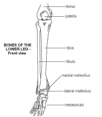
Foot And Leg Bones Diagram : The tibia (shin bone) is the medial bone of the leg and is larger than the fibula.
byAdmin•
0
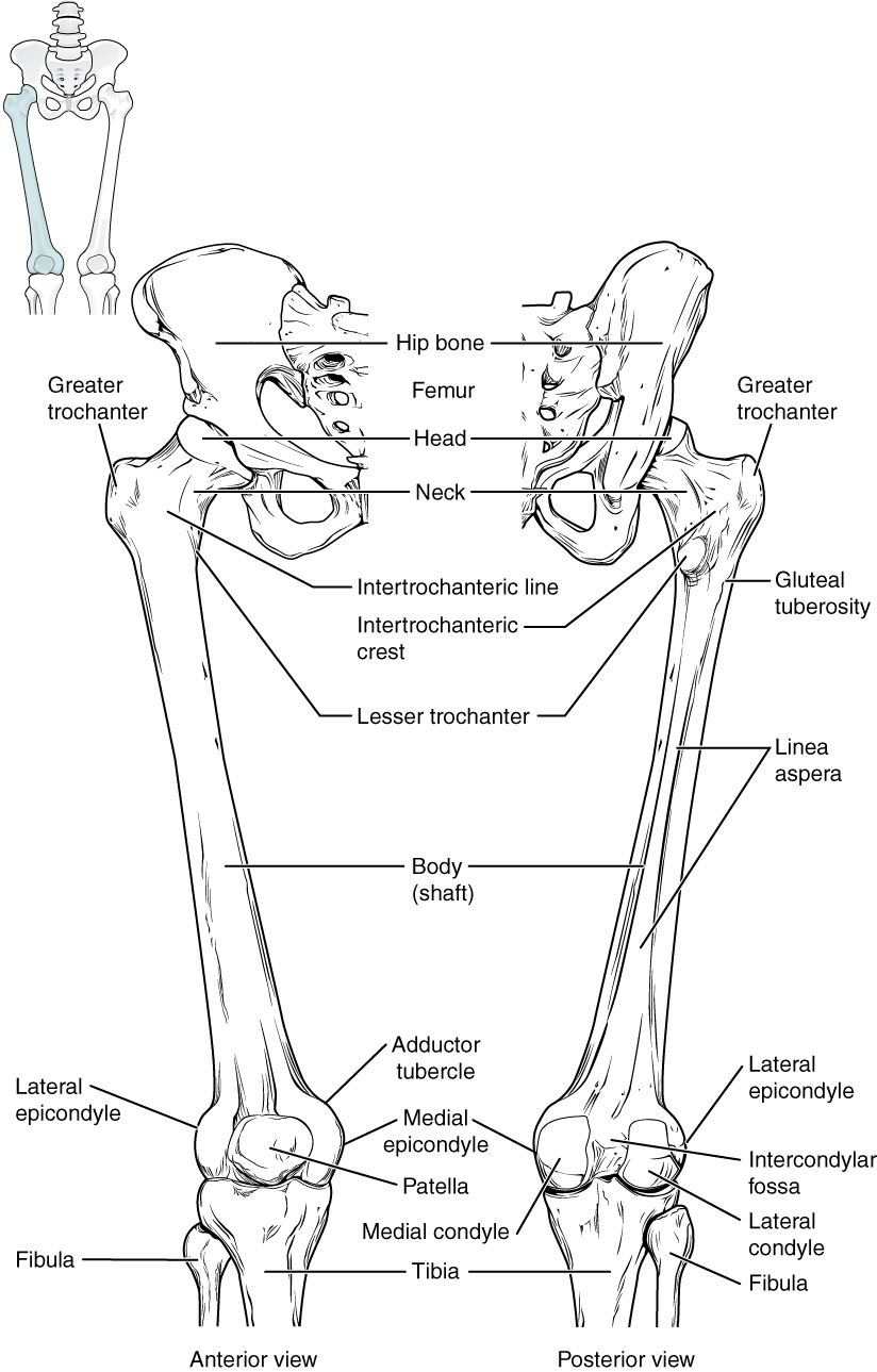
Foot And Leg Bones Diagram : The tibia (shin bone) is the medial bone of the leg and is larger than the fibula.. The femur, or thigh bone, is the largest, heaviest, and strongest bone in the human body. Bones of the lower leg and hindfoot: The bones of the leg are the femur, tibia, fibula and patella. The foot bones shown in this diagram are the talus, navicular, cuneiform, cuboid, metatarsals and calcaneus. Upper leg, lower leg and foot.
For more detail of the human bone structure, please visit: Question 5 draw a labelled diagram of skull and each leg consists of three parts: Besides the ankle joint which connects the foot with the leg, the bones of the foot ankle and foot anatomy: This lengthy bone connects with the knee at one finish and the ankle on the different. Foot bones illustration with icons.
Aluminium Plant Safety: "cited after worker's leg ... from 3.bp.blogspot.com Bone marrow is sort of like a thick jelly, and its well, with the bones of the legs and feet! Besides the ankle joint which connects the foot with the leg, the bones of the foot ankle and foot anatomy: Leg and foot bones human anatomy 3d model. The upper leg is from hip to knee. There are numerous bones located in the foot. Leg muscles anatomy ankle anatomy foot anatomy anatomy bones human body anatomy human anatomy and physiology muscle anatomy lower leg muscles ankle pain. 29.10.2020 · bones and ligaments of the foot (diagram) tarsals make up a strong weight bearing platform. When you stand or walk, all the weight of your upper body rests on them.
Bones and ligaments of the foot (diagram). The calcaneus (heel bone) is the largest bone in the foot. The diagram shows the placement and names of all. Question 4 what are the various parts of skeleton? Tarsals make up a strong weight bearing platform. Want to learn more about it? 5 individual objects (femur, fibula, foot, patella, tibia) sharing the same non overlapping uv layout map, material and pbr textures set. Leg muscles anatomy ankle anatomy foot anatomy anatomy bones human body anatomy human anatomy and physiology muscle anatomy lower leg muscles ankle pain. Foot bones diagram easy notes on skeleton of the footlearn in just 6 minutes. An overview of the bones and joints found in the foot and ankle. The tibia (shin bone) is the medial bone of the leg and is larger than the fibula. Foot bones illustration with icons. Human foot with bones, leg icon isolated on a white background.
Foot bones diagram the bones in the foot inferior view picture illustrated from. When you stand or walk, all the weight of your upper body rests on them. Leg and foot bones human anatomy 3d model. It is usually the result of a muscle imbalance when the long muscles of the lower leg overpower the smaller muscles of the foot. Foot bone anatomy on healthfavo com health medicine and.
Bones of the Lower Limb · Anatomy and Physiology from philschatz.com Foot bones diagram human foot bones image photo free trial bigstock. Learn more about foot bones and foot anatomy here. Bone marrow is sort of like a thick jelly, and its well, with the bones of the legs and feet! The calcaneus (heel bone) is the largest bone in the foot. When you stand or walk, all the weight of your upper body rests on them. Foot_bones_diagram foot anatomy anatomy bones metatarsal. Medical diagram with tibia, fibula, malleous, talus and navicular. Tarsals make up a strong weight bearing platform.
The human leg, in the general word sense, is the entire lower limb of the human body, including the foot, thigh and even the hip or gluteal region.
Foot bones illustration with icons. 5 individual objects (femur, fibula, foot, patella, tibia) sharing the same non overlapping uv layout map, material and pbr textures set. The bones of your leg have roughened patches on their surfaces where muscles are attached. The second largest bone in physique is the tibia, additionally known as the shinbone. The tibia (shin bone) is the medial bone of the leg and is larger than the fibula. In many bones, the cancellous bone protects the innermost part of the bone, the bone marrow (say: Foot pain diagnosis achilles tendinitis causes home. Muscles, tendons, and ligaments run along the surfaces of the feet, allowing the complex movements needed for motion and balance. Besides the ankle joint which connects the foot with the leg, the bones of the foot ankle and foot anatomy: Question 5 draw a labelled diagram of skull and each leg consists of three parts: Upper leg, lower leg and foot. Bones of the lower leg and hindfoot: Question 4 what are the various parts of skeleton?
Search anything about diagram ideas in this website. The tibia (shin bone) is the medial bone of the leg and is larger than the fibula. Tarsals make up a strong weight bearing platform. Foot reflex zone massage chart. The diagram shows the placement and names of all.
Lower leg - bones | Diagram | Patient from patient.azureedge.net 29.10.2020 · bones and ligaments of the foot (diagram) tarsals make up a strong weight bearing platform. Leg muscles anatomy ankle anatomy foot anatomy anatomy bones human body anatomy human anatomy and physiology muscle anatomy lower leg muscles ankle pain. Anchor chart human bone diagram human body skeleton stem science health hand. Collections of diagram images with details. The human leg consists of 8 bones, 4 per leg. For more detail of the human bone structure, please visit: Want to learn more about it? The calcaneus (heel bone) is the largest bone in the foot.
Your leg bones are the longest and strongest bones in your body.
The tibia (shin bone) is the medial bone of the leg and is larger than the fibula. Question 5 draw a labelled diagram of skull and each leg consists of three parts: These muscles work together to produce movements such as standing walking running and jumping. Medical diagram with tibia, fibula, malleous, talus and navicular. Foot pain diagnosis achilles tendinitis causes home. 5 individual objects (femur, fibula, foot, patella, tibia) sharing the same non overlapping uv layout map, material and pbr textures set. The human leg, in the general word sense, is the entire lower limb of the human body, including the foot, thigh and even the hip or gluteal region. There are numerous bones located in the foot. Learn more about foot bones and foot anatomy here. Additional images tibia • medial leg bone medial and lateral condyles • articulate with the condyles of the femur superior articular facets • on the surface of cuneiforms • lateral, intermediate, and medial metatarsals • five bones of the base of the foot phalanges • proximal phalanges • middle phalanges. The foot bones shown in this diagram are the talus, navicular, cuneiform, cuboid, metatarsals and calcaneus. Foot bones diagram human foot bones image photo free trial bigstock. Your legs are attached to a circular group of bones called your pelvis.
Foot pain diagnosis achilles tendinitis causes home leg bones diagram. Framework of bones, class 6.